
门: 未定
纲: 未定
目:开腔骨目
科: 开腔骨科
属:异射骨属
种: 二街异射骨
描述:
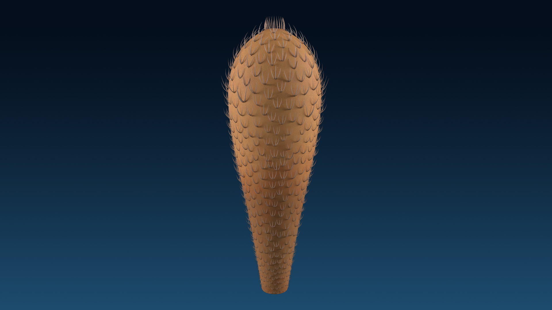
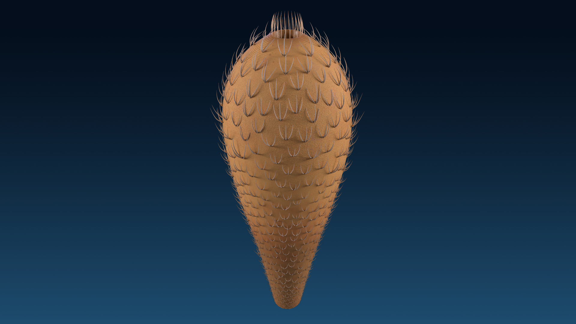
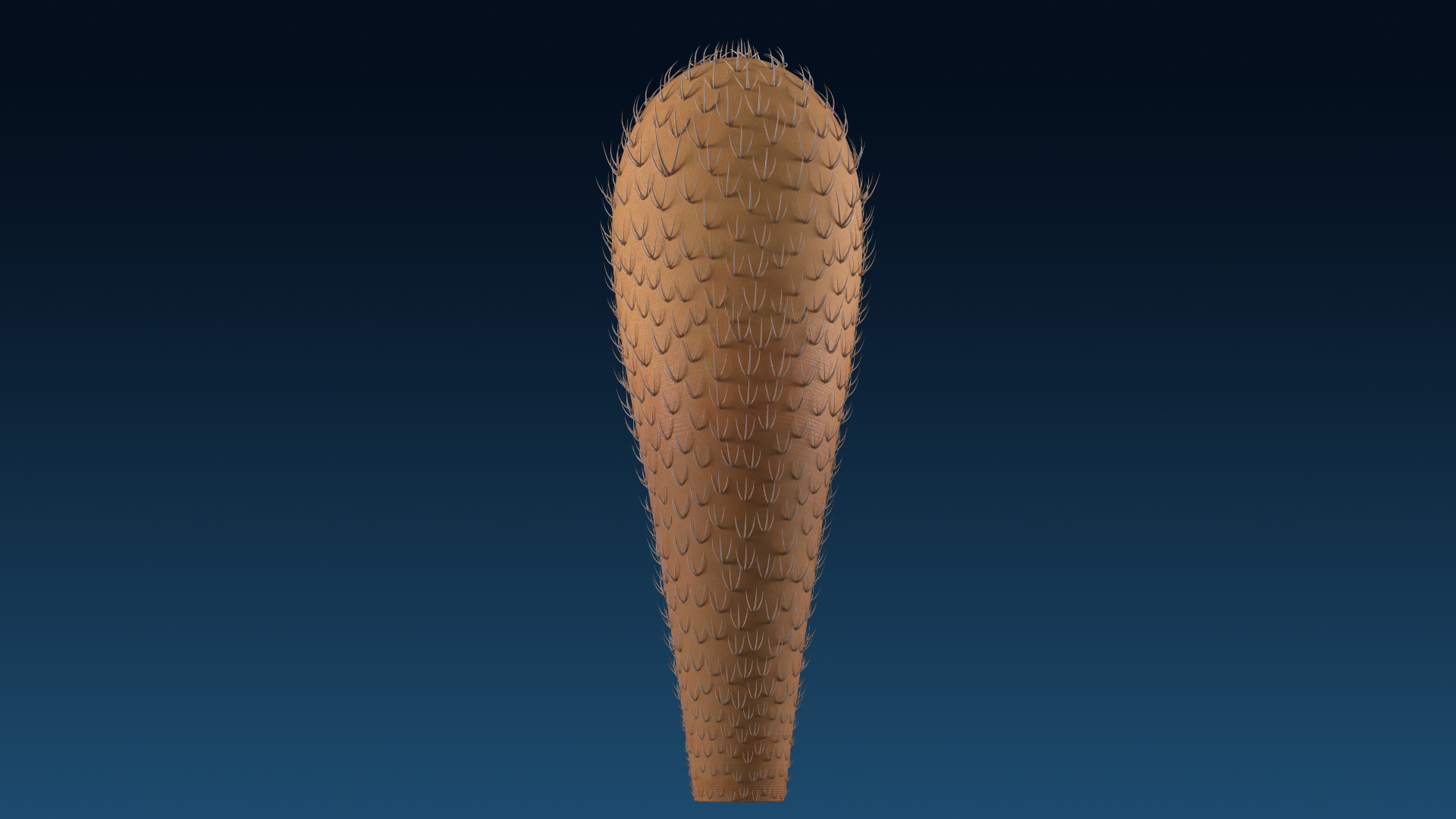
二街异射骨身体呈圆锥形,体表覆盖着数十到数百个三射骨片(通常有两个射保存),让人联想到多刺的火炬。身体的下部逐渐变细,形成钝的基端,上部逐渐加宽,并在顶端迅速收缩。顶端的孔口被密集的单射骨片包围,形成类似的栅栏簇。高约120毫米,基部宽约5毫米,最宽部分35毫米。中间高度处的强烈扭曲和顶点的缺失表明其原始高度超过150 mm。
骨片:体表的骨片通常呈叉形,基盘上有两个可见的侧向射骨,其上通常保留一个圆形突起,对应于第三条射的近端。虽然第三条射骨由于其向外指向的位置很容易被破坏,但在一些标本的骨片中发现了一些典型的三射骨片。明显致密的骨片从体表边缘突出,并向上指向,射与体表之间的角度为60-80°。通常沿射纵轴出现一条薄槽或脊,表明空心射掩埋后压实变形。
在基端,每个骨片的射长1.5–2.5 mm,近身体部较宽的(约0.3 mm宽),向远端急剧变细,形成锐三角形。基端往上,骨片的射明显变大(保守估计为2-3毫米长,最宽处为0.3-0.4毫米宽)。身体中间部分为带有相对纤细的射的骨片,其长3.5–4毫米,宽0.4毫米。在身体的上部,骨片都以红色突起和凹槽的形式保存下来,典型的纤细射长4-5毫米,宽0.5毫米。
表皮:由于紧密分布和相互重叠的骨片,表皮的细节几乎看不到。在一些标本中,骨片之间有相互平行且规则排列的横纹。条纹宽约40-50微米,间距约20微米,可能代表表皮的微小褶皱或装饰。
Phylum: uncertain
Class: uncertain
Order:Chancelloriida Wallcott, 1920
Family: Chacelloridae Wallcott, 1920
Genus:Allonnia Dore & Reid, 1965
Specie: Allonnia erjiensis sp. nov. 2018
Description:
The overall body of Allonnia erjiensis has a conical shape, covered with dozens to hundreds of three rayed sclerites (usually preserved with two lateral rays), reminiscent of a thorny torch. The lower part of the body tapers abapically to a blunt basal end, and the upper part widens apically, and is rapidly contracted into an apex at the top. The orifice on the apex is surrounded by dense single-rayed sclerites, forming a putative palisade-like tuft. The holotype is about 120 mm high and 5 mm wide at the basal end, broadening apically to 35 mm at its widest part. The strong twist at the middle height and the absence of the apex suggest its original height was over 150 mm.
Sclerites: Sclerites on the body surface are mostly wishbone-like in shape, with two tangible lateral rays articulated in the basal disk, on which a round protrusion is usually preserved corresponding to the proximal part of the third ray (ascending ray). Though the ascending rays are easily broken due to their outwards-pointing position, a few typical three-rayed sclerites are found in the scleri-tome of some specimens. Obvious and dense sclerites protrude from the marginal parts of the body surface and point upwards in which the angle between the ray (possibly the ascending ray) and the body surface is 60–80°. A thin groove or ridge usually presents along the longitudinal axis of the ray, suggesting the deformation of the hollow ray under compaction after entombment.
In the basal end, the rays of each sclerite are 1.5–2.5 mm long and have wide proximal parts (approximately 0.3 mm wide) which taper sharply towards the distal part, forming an acute-triangular shape. In the abapical part of the body, the rays of sclerites are slightly larger (conservatively estimated as 2–3 mm long and 0.3–0.4 mm wide at their widest part). The detail of the marginal area of the body over the twisted middle part, possessing sclerites with relatively gracile rays, which are 3.5–4 mm long and 0.4 mm wide. In the upper part of the body, sclerites are all preserved as reddish bulges and troughs. The typical gracile rays are 4–5 mm long and 0.5 mm wide.
Integument: In general, details of the integument are hardly seen due to the densely distributed and mutually overlapping sclerites. There are gleaming transverse stripes which are parallel to each other and regularly arranged between the sclerites in some specimens. The stripes are approximately 40–50 μm wide and about 20 μm apart, probably representing the tiny folds or ornamentations of the integument.
年龄:寒武纪第二世第三阶
主要产地: 澄江生物群(云南澄江)
Age: Cambrian Series2, Stage3
Principal localities: Chengjiang biota (Yu’anshan Member of the Chiungchussu Heilinpu Formation), Yunnan Province, China
Yun, Hao & Zhang, Xingliang & Li, Luoyang. (2017). Chancelloriid Allonnia erjiensis sp. nov. from the Chengjiang Lagerstätte of South China. Journal of Systematic Palaeontology. 16. 1-10. 10.1080/14772019.2017.1311380.