

门: 节肢动物门
纲: 大附肢纲
目:未定
科:剪刀手虫科
属:剪刀手虫属
种: 德式剪刀手虫
描述:
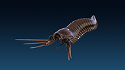
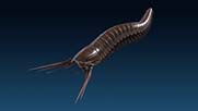
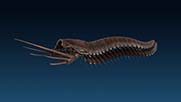
头部长约4mm(为躯干10%长)。头部保存完好,由狭窄的前半部分和扩大的后半部分组成。前半部分有两只带柄的,直径约0.9mm的大复眼。后半部分头甲向腹部弯曲并且至少包含一对大附肢,可能在眼睛周围还有一对触角。头甲后边缘覆盖在第一节躯干体节之上。头甲后半部分很多条纹可能揭示标本活着的时候该部分是弯曲的。触角保存较差,只能看到细丝状的点缀,点缀之间可能代表了触角是分节的。大附肢由近端的茎和三节远端带刺的肢节组成。最近一节带有最长的刺(长约11.1mm,占附肢总长的70%),但是它没有其他刺强壮且轻微弯曲。位于前部的肠道保存较好,揭示了标本有一个腹侧的嘴巴。中肠腺从肠道延伸至头甲后部及躯干上,由三或四个放射指状突起组成。其至少延申至第六体节,8-15节之间有零碎的肠道组织保存下来。躯干约43.5mm长且由29节体节组成,体节向后逐渐变窄。每一节体节都只有一对附肢。尾节不明显。
Phylum: Arthropoda
Class: Megacheira Order:
Order:uncertain
Family: Kootenichelidae
Genus:Kootenichela
Specie: Kootenichela deppi
Description:
The cephalon is 4 mm long (,10% preserved body length),measured from the anterior-most margin to the postero-dorsal margin.This is best preserved in the part and consists of two distinct regions: a narrow anterior and an expanded posterior.The anterior region bears two large pedunculate eyes 0.9 mm in diameter; a small rounded stain within the cephalon is the right eye and appears to preserve individual lenses.Individual lenses can also be distinguished on the left eye and number 50/mm2.The posterior margin of the cephalon curves antero-ventrally and encompasses at least the ‘‘great-appendages’’ and possibly an antenna-like appendage on the ocular segment. There are no other cephalic limbs although the posterior of the cephalon appears to overlap the first trunk tergites, which led to the previous misinterpretation that it possessed four-limb bearing segments.Numerous striations in the posterior part of the cephalon may indicate that it was convex in life and was crushed post-mortem. The putative antenna is preserved as a narrow, almost filamentous staining with little structural detail. This structure is interpreted as a true
biological structure rather than abiogenic staining, which is not found elsewhere in material from this horizon. Gaps in the staining may represent antennal segments. The ‘‘great-appendages’’ are composed of a bipartite proximal peduncle and three distal
spine-bearing podomeres.Although poorly preserved in the holotype, the spines have similar proportions and degree of recurvature to those in a well-preserved isolated pair of cooccurring ‘‘great-appendages’’ that are herein considered conspecific. The most proximal spinebearing podomere has the longest spine, 11.1 mm in length (70% total appendage length), less robust than the others and slightly recurved.An anterior gut trace is preserved;this documents a ventrally directed mouth and extends into the anterior cephalic region , where the dark staining may result from gut-rupture.Midgut glands extend from the gut into the posterior cephalon and trunk and consist of tri-or tetra-radiate finger-like projections. They extend posteriorly to (at least) segment 6, and a strand of unbranched gut is preserved between segments 8 and 15.The preserved part of the trunk in the holotype is 43.5 mm long and consists of 29 segments,tapering posteriorly. Each segment bears a single pair of limbs.
时代:寒武系第三统第五阶,布尔吉斯页岩组
主要产地: 加拿大不列颠哥伦比亚省约霍国家公园,布尔吉斯生物群
Age:Cambrian (Series 3, Stage 5), Burgess Shale Formation
Principal localities: Burgess biota (in Yoho National Park,British Columbia,Canada)
David Legg,2013.Multi-Segmented Arthropods from the Middle Cambrian of British Columbia (Canada).Paleontology,Volume 87 Issue 3,pp.493 - 501.
Cédric Aria,2020.Fossils from South China redefine the ancestral euarthropod body plan.BMC Evolutionary Biology,Volume 20, Article number: 4.